Anatomy Of Chest Area / Anatomy and Physiology of the Lungs | Health Life Media - The epidermis is the outermost layer that provides a protective, waterproof seal over the body.
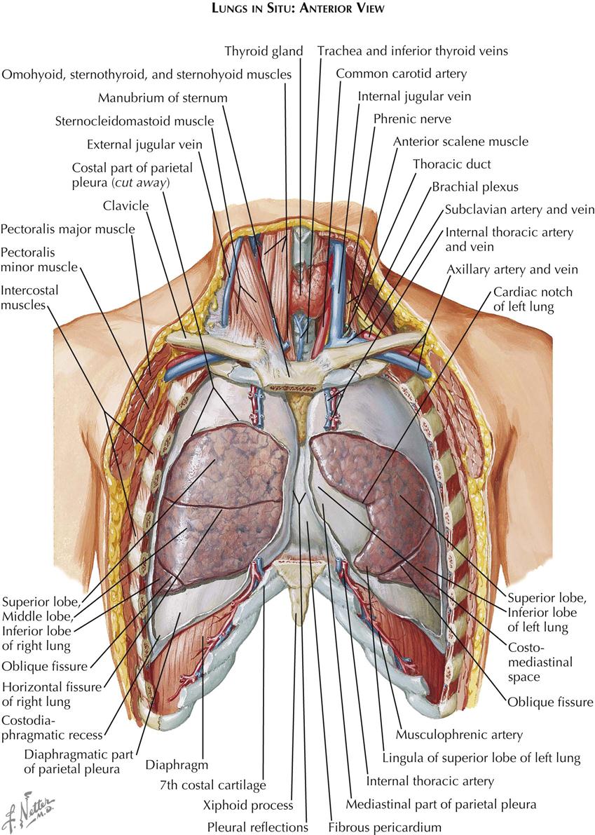
Anatomy Of Chest Area / Anatomy and Physiology of the Lungs | Health Life Media - The epidermis is the outermost layer that provides a protective, waterproof seal over the body.. Anatomy of the chest and the lungs: 1, inferior lobe of right lung. Iv contrast may be injected into a vein in the patient's arm or hand. These areas are also known as the hidden areas. Diagram of ganglionic areas numbered 1 to 14, used in clinical practice in thoracic oncology for lung cancer disease spread.
Ct anatomy of the chest, axial reconstruction. In insects, crustaceans, and the extinct trilobites, the thorax is one of the three main divisions of the creature's body. 1, inferior lobe of right lung. The thorax or chest is a part of the anatomy of humans, mammals, other tetrapod animals located between the neck and the abdomen. Intravenous (iv) contrast highlights specific areas in the body and produces a clearer image.
1. Anatomy | Thoracic Key from thoracickey.com You can support the work of campbellteaching, at no cost whatsoever to yourself, if you use the link below as your bookmark to access amazon. 1, inferior lobe of right lung. Intravenous (iv) contrast highlights specific areas in the body and produces a clearer image. These areas are also known as the hidden areas. Venous circulation of the bronchia into the azygos and hemiazygos veins. Anatomy of the chest and the lungs: In insects, crustaceans, and the extinct trilobites, the thorax is one of the three main divisions of the creature's body. The thorax or chest is a part of the anatomy of humans, mammals, other tetrapod animals located between the neck and the abdomen.
In insects, crustaceans, and the extinct trilobites, the thorax is one of the three main divisions of the creature's body.
Hilar lymph nodes are not visible unless abnormal. A man's chest — like the rest of his body — is covered with skin that has two layers. In insects, crustaceans, and the extinct trilobites, the thorax is one of the three main divisions of the creature's body. Anatomy of the chest and the lungs: Pathology of the heart, mediastinum, lungs and pleura. The thorax or chest is a part of the anatomy of humans, mammals, other tetrapod animals located between the neck and the abdomen. You can support the work of campbellteaching, at no cost whatsoever to yourself, if you use the link below as your bookmark to access amazon. Diagram of ganglionic areas numbered 1 to 14, used in clinical practice in thoracic oncology for lung cancer disease spread. Each hilum contains major bronchi and pulmonary vessels. These areas are also known as the hidden areas. Iv contrast may be injected into a vein in the patient's arm or hand. 1, inferior lobe of right lung. Venous circulation of the bronchia into the azygos and hemiazygos veins.
In insects, crustaceans, and the extinct trilobites, the thorax is one of the three main divisions of the creature's body. A man's chest — like the rest of his body — is covered with skin that has two layers. Pathology of the heart, mediastinum, lungs and pleura. Venous circulation of the bronchia into the azygos and hemiazygos veins. Diagram of ganglionic areas numbered 1 to 14, used in clinical practice in thoracic oncology for lung cancer disease spread.
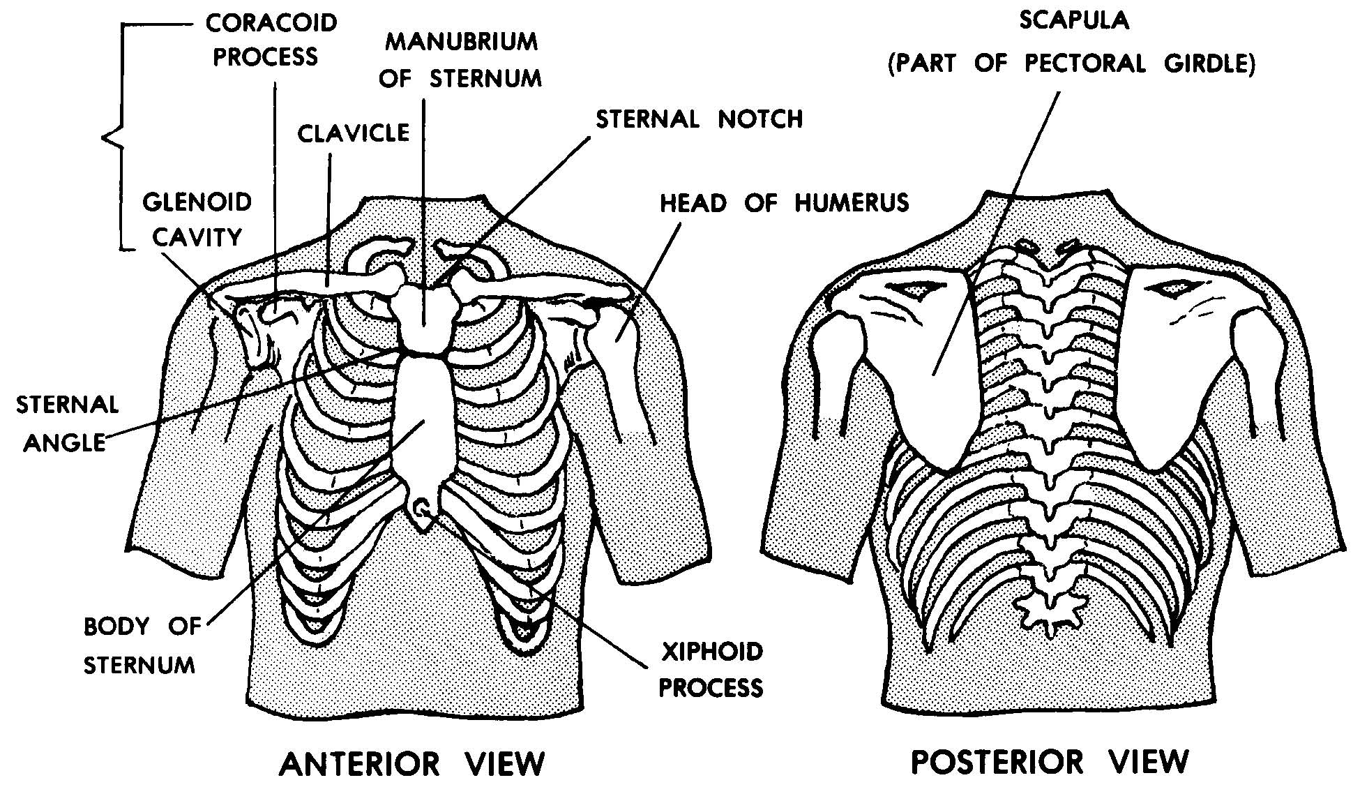
Images 04. Skeletal System | Basic Human Anatomy from brooksidepress.org Venous circulation of the bronchia into the azygos and hemiazygos veins. Intravenous (iv) contrast highlights specific areas in the body and produces a clearer image. Hilar lymph nodes are not visible unless abnormal. Ct anatomy of the chest, axial reconstruction. These areas are also known as the hidden areas. 1, inferior lobe of right lung. The epidermis is the outermost layer that provides a protective, waterproof seal over the body. Anatomy of the chest and the lungs:
A man's chest — like the rest of his body — is covered with skin that has two layers.
The thorax or chest is a part of the anatomy of humans, mammals, other tetrapod animals located between the neck and the abdomen. These areas are also known as the hidden areas. Anatomy of the chest and the lungs: You can support the work of campbellteaching, at no cost whatsoever to yourself, if you use the link below as your bookmark to access amazon. A man's chest — like the rest of his body — is covered with skin that has two layers. Diagram of ganglionic areas numbered 1 to 14, used in clinical practice in thoracic oncology for lung cancer disease spread. Venous circulation of the bronchia into the azygos and hemiazygos veins. Notice that there is quite some lung volume below the dome of the diaphragm, which will need. Each hilum contains major bronchi and pulmonary vessels. 1, inferior lobe of right lung. Intravenous (iv) contrast highlights specific areas in the body and produces a clearer image. The epidermis is the outermost layer that provides a protective, waterproof seal over the body. Pathology of the heart, mediastinum, lungs and pleura.
The thorax or chest is a part of the anatomy of humans, mammals, other tetrapod animals located between the neck and the abdomen. 1, inferior lobe of right lung. Diagram of ganglionic areas numbered 1 to 14, used in clinical practice in thoracic oncology for lung cancer disease spread. Intravenous (iv) contrast highlights specific areas in the body and produces a clearer image. Each hilum contains major bronchi and pulmonary vessels.
How Do the Bones in My Corset Affect the Bones in My Body? from i1.wp.com Pathology of the heart, mediastinum, lungs and pleura. You can support the work of campbellteaching, at no cost whatsoever to yourself, if you use the link below as your bookmark to access amazon. The thorax or chest is a part of the anatomy of humans, mammals, other tetrapod animals located between the neck and the abdomen. Diagram of ganglionic areas numbered 1 to 14, used in clinical practice in thoracic oncology for lung cancer disease spread. 1, inferior lobe of right lung. Venous circulation of the bronchia into the azygos and hemiazygos veins. Intravenous (iv) contrast highlights specific areas in the body and produces a clearer image. Iv contrast may be injected into a vein in the patient's arm or hand.
The epidermis is the outermost layer that provides a protective, waterproof seal over the body.
Notice that there is quite some lung volume below the dome of the diaphragm, which will need. Ct anatomy of the chest, axial reconstruction. Pathology of the heart, mediastinum, lungs and pleura. Iv contrast may be injected into a vein in the patient's arm or hand. Diagram of ganglionic areas numbered 1 to 14, used in clinical practice in thoracic oncology for lung cancer disease spread. 1, inferior lobe of right lung. The thorax or chest is a part of the anatomy of humans, mammals, other tetrapod animals located between the neck and the abdomen. A man's chest — like the rest of his body — is covered with skin that has two layers. These areas are also known as the hidden areas. Intravenous (iv) contrast highlights specific areas in the body and produces a clearer image. The epidermis is the outermost layer that provides a protective, waterproof seal over the body. In insects, crustaceans, and the extinct trilobites, the thorax is one of the three main divisions of the creature's body. Hilar lymph nodes are not visible unless abnormal.
Notice that there is quite some lung volume below the dome of the diaphragm, which will need anatomy of chest. You can support the work of campbellteaching, at no cost whatsoever to yourself, if you use the link below as your bookmark to access amazon.